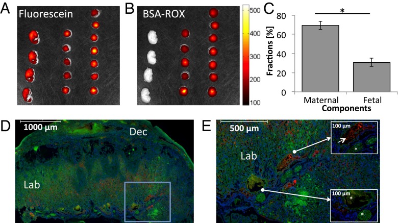
Fig. 6.
Optical imaging of maternal and fetal blood fractions on ICR pregnant mice. (A and B) Fluorescence intensity analyzed using an IVIS imaging system following the injection of fluorescein (A) and BSA-ROX (B). These ex vivo analyses involved n = 6 dams and 27 placentas/fetuses. (C) Estimated maternal and fetal blood fractions derived for a cohort of placentas from this optical microscopy analysis (mean ± SEM; P < 0.05). (D and E) Representative fluorescence images of placental histology sections, showing the circulatory bed of the placental labyrinth. BSA-ROX (red) was confined to the maternal circulation within the labyrinth, whereas fluorescein (green) also penetrated the fetal circulation. Markers in D and E represent 1,000 µm and 500 µm, respectively. Blue frame in D is magnified in E, whereas Insets in E indicate the localization of a maternal blood pool (Upper Right; marker indicates 100 μm) as well as fetal blood spaces (Lower Left; asterisks in Insets mark the fetal blood spaces and marker indicates 100 μm). Blue: Hoechst-stained nuclei. Dec, decidua; Lab, labyrinth.

