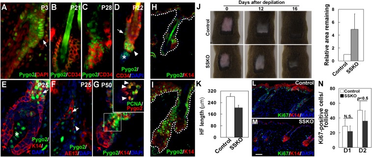
Fig. 1.
Pygo2 is dynamically expressed in HF SC/EPCs, and it facilitates depilation-induced hair regeneration. (A–G) Indirect immunofluorescence of HFs at different postnatal (P) ages using the indicated antibodies. DAPI stains the nuclei. White arrows indicate bulge cells in A and D, K14+ ORS cells in E, and AE13+ precortex cells in F. The red arrow in E and G indicates matrix cells. White arrowheads indicate HG cells in D, IRS cells adjacent to the precortex in F, and Pygo2+/PCNA+ cells in G. White asterisks in D and E denote Pygo2+ DP cells. Inset in G shows an enlarged image of the boxed area. (H and I) Hair depilation up-regulates Pygo2 protein. Control (H) and depilated (I) skin of the same mouse were analyzed 2 d after depilation (D2). (J) Loss of Pygo2 delays postdepilation hair regeneration. For quantitative analysis (Right), the area that remained in each mouse (n = 5 for control; n = 3 for SSKO) was measured at D11–16, and that of the control was arbitrarily set to be 1. (K) Reduced HF length in depilated SSKO skin at D2 (n = 3). (L–N) Ki67 staining of control and SSKO skin at D1 and D2. Quantification (N) was done for proliferative cells in the HF (n = 3). Error bars denote standard deviation (SD). Control genotypes, Pygo2+/f or K14-Cre/Pygo2+/f. [Scale bar, 10 μm (A–G), 15 μm (H and I), and 45 μm (L and M).]

