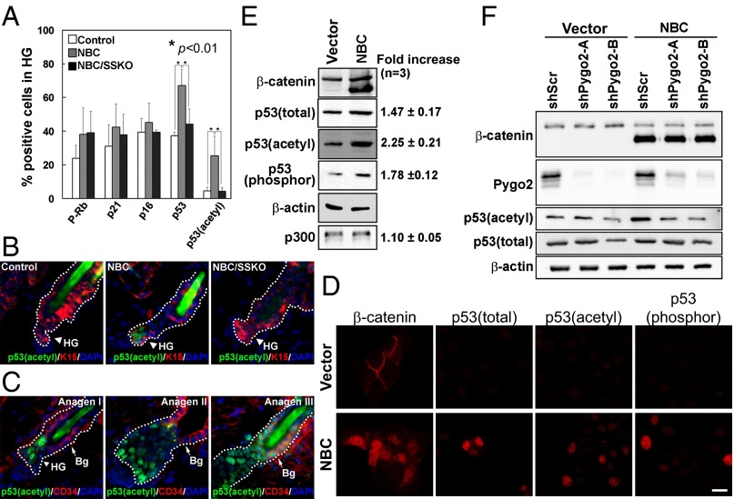
Fig. 4.
Pygo2-dependent p53 activation upon β-catenin overexpression and T–A transition. (A) Quantitative analysis for cell-cycle regulators in telogen HFs from control, NBC, and NBC/SSKO mice. Error bars denote SD. (B) Representative images showing acetyl-p53 immunostaining results. Arrowhead and arrow indicate K15+ HG and CD34+ bulge cells, respectively. See Fig. 2 legends for control genotypes. (C) Emergence of acetyl-p53 during normal T–A transition. (D and E) Indirect immunofluorescence (D) and Western blotting (E) of various forms of p53 protein in HaCaT cells. Values on the right indicate β-catenin–induced fold change. (F) Effect of Pygo2 depletion on p53 protein. [Scale bar, 20 μm (B and C) and 10 μm (D).]

