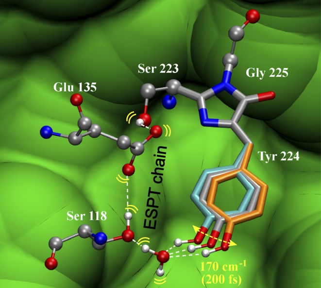
Fig. 5.
Illustration of initial structural evolution gating ESPT in the Ca2+-free GEM-GECO1. Geometric constraints from the Ca2+-free GCaMP2 crystal structure 3EKJ are used. An optimized ESPT pathway is delineated with an intervening H2O molecule added (Fig. S4B) due to limited resolution of the crystal structure. To obtain an SYG chromophore, the methyl group of T223 is replaced by an H atom without further structural modification. The 170 cm−1 mode facilitates ESPT with three positions depicted (DFT-calculated mode displacements are ±2° in equilibrated S0). The bridge C—C single bond shifts from the inherent structure (gray, 125°, the bridge C = C–C angle) back (orange, 132°) and forth (cyan, 118°) to modulate H-bonding geometry in S1 with the adjacent H2O molecule. C, O, N, and H atoms are shown in gray, red, blue, and white, respectively. The surrounding protein environment is shown in green. Rendered using visual molecular dynamics (VMD) (49).

