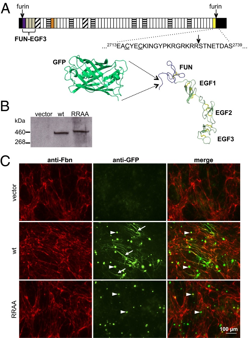
Fig. 1.
GFP-Fbn fusion design and microfibril binding assay. (A) The structure of fibrillin-1 is dominated by calcium-binding EGF-like domains (white) interspersed with transforming growth factor β-binding protein-like (horizontal stripes) and hybrid (diagonal stripes) domains. Other regions include the fibrillin unique N-terminal (FUN; purple) and non-calcium-binding EGF-like (gray) domains, a proline-rich region (orange) and a conserved 2Cys domain (yellow). N- and C-terminal propeptides (black) are processed at furin cleavage sites (arrows) before microfibril assembly. The sequence around the C-terminal furin cleavage site is shown in relation to the cysteines (underlined) of the 2Cys domain. A model of GFP (pdb 2YOG), drawn to scale next to the fibrillin N-terminal domains (FUN-EGF3; pdb 2M74), is provided to show the size of the tag relative to the fibrillin-1 N terminus. (B) Anti-GFP Western blot of medium samples from cultures of HEK293T cells transiently transfected with empty vector or constructs encoding either GFP-Fbn or the GFP-FbnRRAA mutant (RRAA). The RRAA variant is secreted and has the expected shift to a higher molecular weight, confirming a lack of C-terminal processing. (C) FS2 fibroblasts cocultured with HEK293T cells transiently transfected with either empty vector (pcDNA), pcDNA-GFPFbn (GFP-Fbn) or pcDNA-GFPFbnRRAA (RRAA). Cells were stained for fibrillin (red) and GFP (green). GFP-Fbn cocultures showed extensive GFP-positive fibrillar networks (arrows). Although the RRAA mutant was secreted into the medium at levels comparable to wild-type GFP-Fbn (B), no GFP-positive fibrillar network was observed in RRAA cocultures. Intracellular GFP fluorescence (arrowheads) suggested similar transfection efficiencies in the GFP-Fbn and RRAA experiments. These cells were not stained with anti-fibrillin antibody under the conditions used, suggesting that the green fluorescence was due to intracellular accumulations of recombinant protein. This was verified by comparing the anti-fibrillin staining of transiently transfected HEK293T cells in single culture with or without permeabilization (Fig. S1).

