
Figure 2. Amino acid sequence and membrane topology of human GLUT1.
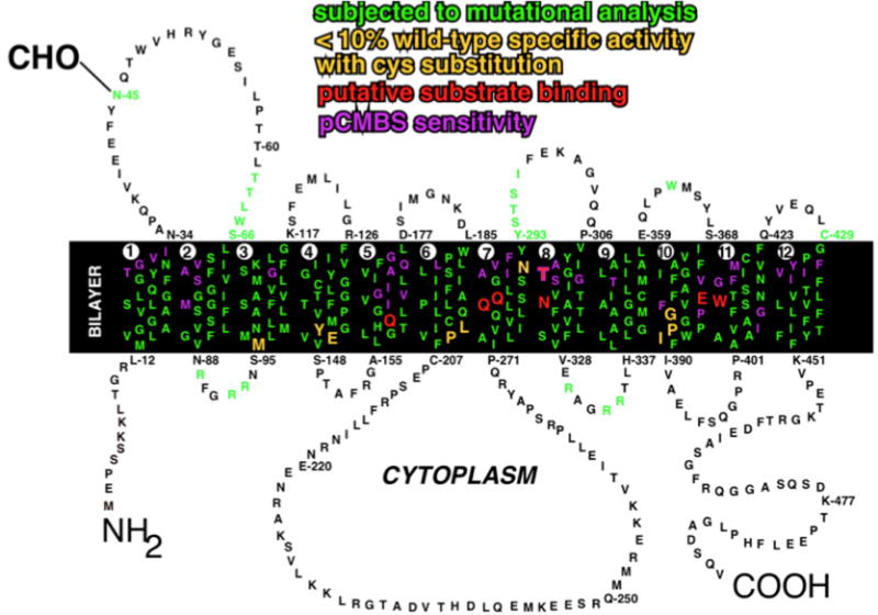
Amino acid residues are designated by the single letter code. The transmembrane segments are numbered 1–12. The linkage of the N-linked oligosaccharide at N45 is shown. Positions that have been analyzed by site-directed mutagenesis are in green. Residues that are believed to play a direct role in substrate binding are shown in red. Residues that are accessible to pCMBS from the external solvent are shown in purple. Residues that appear to be critical for transport activity are shown in yellow. Adapted from (Mueckler and Makepeace, 2009).