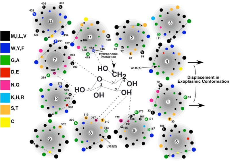
Figure 3. Model of the exoplasmic substrate-binding site of GLUT1 (Mueckler and Makepeace, 2009).

Glucose is not drawn to scale. The arrangement of helices is shown in a simplistic fashion for clarity. Amino acid residues that are in contact with solvent in the aqueous cavity are numbered and identified by the single-letter code. Dotted lines represent putative hydrogen bonds.
