
Figure 5. Colocalization of GFP-tagged wild type GLUT4 and HA-tagged IRM mutant GLUT4 by immunofluorescence laser confocal microscopy.
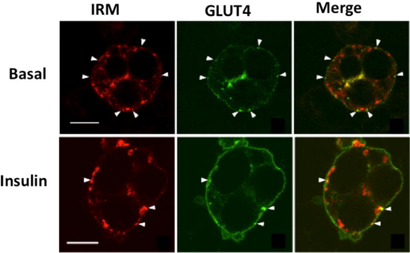
3T3L1 adipocytes were co-infected with recombinant adenoviruses encoding the GFP-tagged wild-type GLUT4 and a HA-tagged IRM mutant (see text). 48 h later the cells were serum-starved for two hours, and then either exposed to insulin for 30 min or maintained in the basal state. Tagged wild-type GLUT4 is shown in the middle panels in green, the tagged mutants are shown in the left panels in red, and the merged images are shown in the right panels with colocalization between the two coexpressed proteins presented in yellow. The scale bars represent 10 μM. Note the redistribution of wild-type GLUT4 to the plasma membrane after insulin treatment and the lack of redistribution in the IRM mutant. Additionally, the mutant and wild type transporter are largely present in distinct intracellular membranes in the absence or presence of insulin. Adapted from (Song et al., 2008).