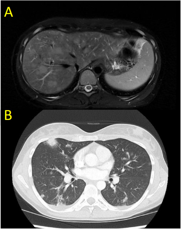
Figure 1.

Diagnostic imaging features of lymphomatoid granulomatosis in our case. A) T2 weighted axial magnetic resonance (MR) image showing hyperintense lesions in the liver. B) CT axial image showing irregular bilateral pulmonary opacities with peripheral predominance and areas of ground-glass changes classic for lymphomatoid granulomatosis.
