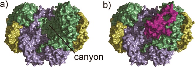
Figure 1.

Crystal structure of the hydroxylase–regulatory protein complex of sMMO (PDB ID 4GAM): (a) the hydroxylase MMOH showing the canyon, and (b) MMOH in complex with the regulatory protein MMOB. There is another MMOB molecule binding to the canyon on the other side of MMOH. MMOH α-subunit is colored in green, β-subunit in blue, γ-subunit in yellow, and MMOB in purple.
