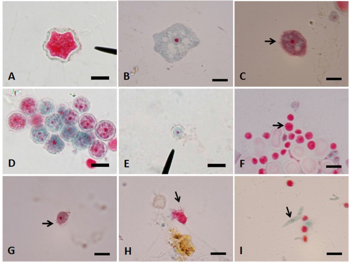
Fig. 2. A-I - Trichrome staining of free-living amoebae under light microscopy (1,000x), bar = 10 µm. A: A cyst of Acanthamoeba sp. B: A large trophozoite of Naegleria sp. C: Acanthamoeba-like trophozoites (arrow showing fine short acanthopodia). D, E: Different sizes of unidentified double-walled cysts with distinct nuclei. F: Unidentified amoebae with small round cysts (arrow). G, H: Small Echinamoeba-like trophozoites (arrow showing few spiny short pseudopodia). I: An elongated cylindrical trophozoite of Hartmannella-like amoeba (arrow).

