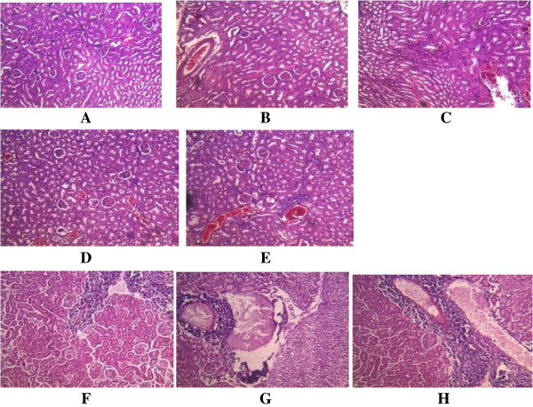
Figure 2.

Histological appearance of the kidney. A. The control, showing no pathological changes. BC. Mice infected with A246A* showing B. essentially unremarkable, normal cells C. minimal infiltration of interstitial by inflammatory cells. DE. Mice infected with A80A* showing D. inflammation from mild to moderate E. infiltration of tubule interstitial, also congestion of vessel and mild compression of capillary luminal. FGH. Mice infected with A245A* showing F. kidney glomerulli with essentially unremarkable cells. G. necrosis H. intense infiltration on the tubule interstitial by chronic inflammatory cells. *As shown in Table 1.
