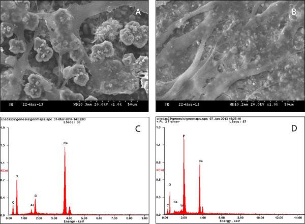
Figure 2.

Investigation of cell morphology and chemical composition of the materials. SEM observation of cells incubated for 3 days on (A) MTA (×1000) and (B) TCP (×1000). Both groups showed flattened cells in close proximity to one another, and these were seen to be spreading across the substrate. EDS analysis of the samples: (C) MTA and (D) TCP.
