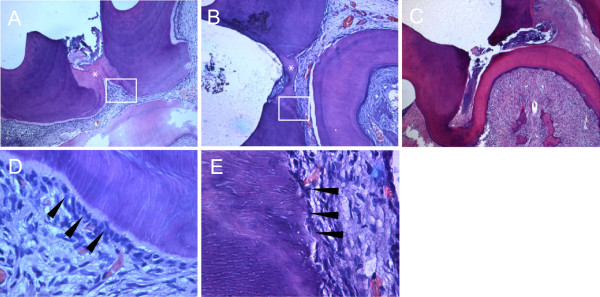
Figure 5.

Histological observation. Capped pulps stained with hematoxylin-eosin 4 weeks after treatment with MTA (A) and TCP (B) (×50). (C) A specimen in the control group capped only with glass ionomer cement. (D and E) Higher magnification of boxed areas shown in A and B (×400), respectively. Odontoblasts (arrowheads) are polarized and appear to be arranged in a palisade pattern. *Reparative tertiary dentin formed underneath the capping materials.
