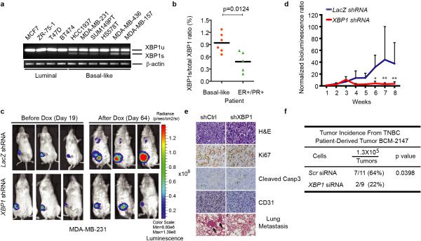
Figure 1. XBP1 silencing blocks TNBC cell growth and invasiveness.
a-b, RT-PCR analysis of XBP1 splicing in luminal and basal-like cell lines (a) or primary tissues from 6 TNBC patients and 5 ER/PR+ patients (b). XBP1u: unspliced XBP1, XBP1s: spliced XBP1. β-actin was used as loading control. c, Representative bioluminescent images of orthotopic tumors formed by MDA-MB-231 cells as in (Extended Data 1d). Bioluminescent images were obtained 5 days after transplantation and serially after mice were begun on chow containing doxycycline (day 19) for 8 weeks. Pictures shown are the day19 image (Before Dox) and day 64 image (After Dox). d, Quantification of imaging studies as in (c). Data are shown as mean ± SD of biological replicates (n=8). *p<0.05, **p<0.01. e. H&E, Ki67, cleaved Caspase 3 or CD31 immunostaining of tumors or lungs 8 weeks after mice were fed chow containing doxycycline. Black arrows indicate metastatic nodules. f, Tumor incidence in mice transplanted with BCM-2147 tumor cells (10 weeks post-transplantation). Statistical significance was determined by Barnard's test22, 23.

