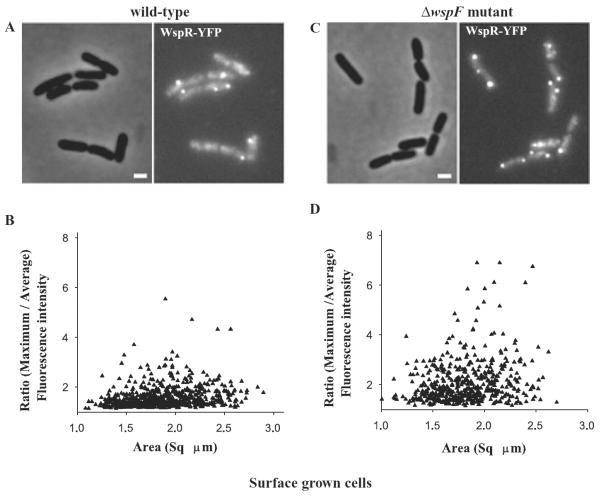
Fig. 7.
Surface growth stimulates WspR–YFP cluster formation in wild-type cells.
WspR–YFP localization in (A) wild-type cells grown on LB agar. The phase-contrast (left) and fluorescence (right) images of cells expressing wspR–yfp are shown.
B. Single cell measurements of fluorescence signal intensities expressed as the ratio of maximum to average signal intensities plotted against the area of the respective single cells in wild-type cells expressing WspR–YFP.
C. ΔwspF mutant cells grown on agar and expressing WspR–YFP.
D. Single cell measurements of fluorescence signal intensities in ΔwspF mutant cells expressing WspR–YFP. Single cell fluorescence data were collected from three independent experiments.

