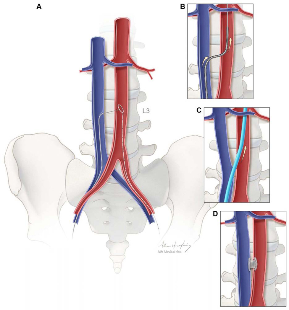
Figure 1. Schematic depiction of caval-aortic access.
(A) A catheter directs a transfemoral vein guidewire from the inferior vena cava towards a snare target positioned in the adjoining abdominal aorta. (B) A catheter is advanced over the guidewire into the aorta and used to introduce a more rigid guidewire. (C) The valve introducer sheath is advanced from the vena cava into the aorta. (D) After completion of TAVR, the aorto-caval access tract is closed with a nitinol occluder.

