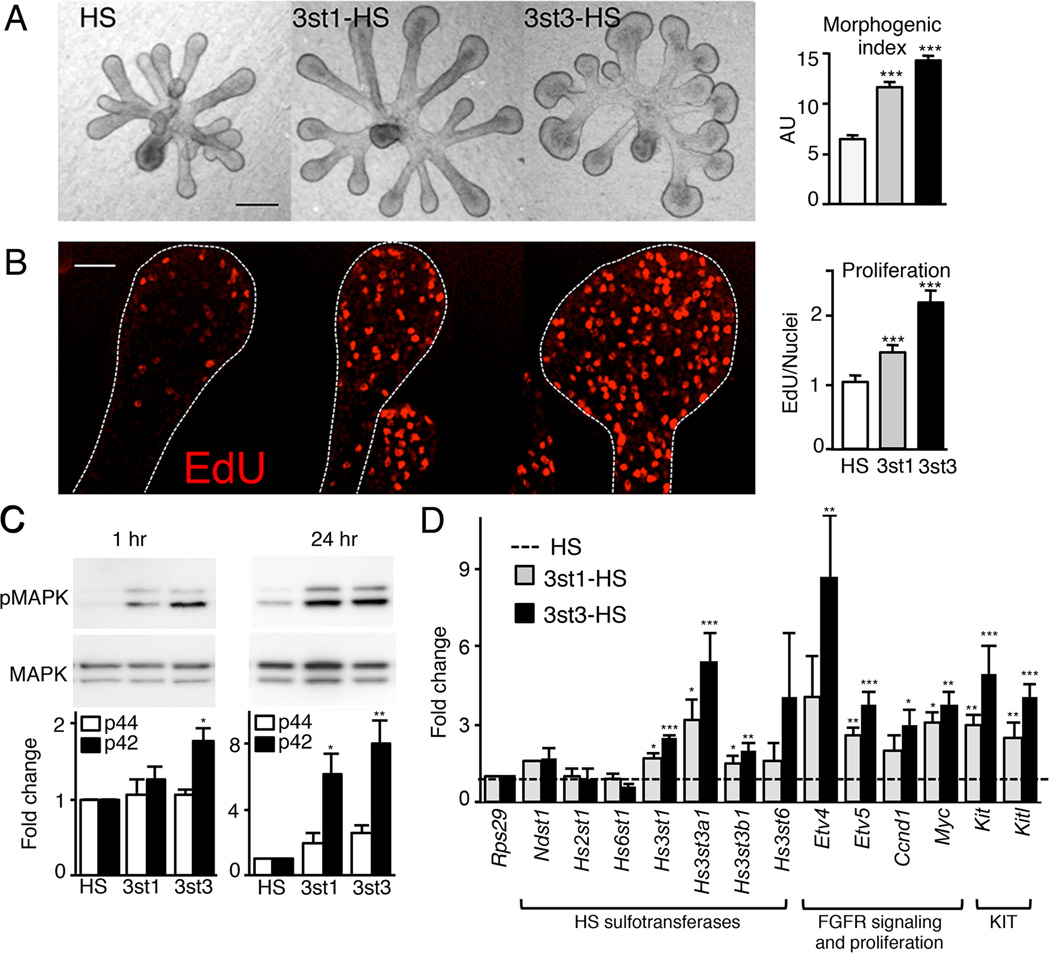
Figure 4. 3-O-sulfated-HS increases FGF10-dependent epithelial morphogenesis by increasing proliferation and FGFR2b-downstream gene expression.
(A) FGF10 cultured E13 SMG epithelia treated with HS, 3st1-HS or 3st3-HS for 28 hr. 3st3-HS increased endbud morphogenesis of FGF10-treated epithelium compared to HS (control). Morphogenic index (endbud number × width × duct length, in AU) increases with addition of 3-O-HS. Scale bar; 50 µm.
(B) Single confocal sections showing cell proliferation (red) in epithelia cultured for 28 hr. Quantification of fluorescence intensity normalized to total nuclei staining and expressed as a ratio to the HS treated epithelia. At least five epithelia/condition were used for quantification from three independent experiments. Scale bar; 10 µm.
(C) 3st3-HS increased MAPK signaling within 1 hr and both 3st-HS maintained it at 24 hr in FGF10-cultured epithelia. Band intensity of pMAPK was normalized to total MAPK and expressed as a fold change in ratio compared to HS. The graphs are 3 experiments combined and the blots from a representative experiment.
(D) Gene expression was measured by qPCR and normalized to epithelia cultured for 28 hr with HS and Rps29.
Error bars: SEM. ANOVA; *p < 0.05; **p < 0.01, and ***p < 0.001.
See also Figure S3.

