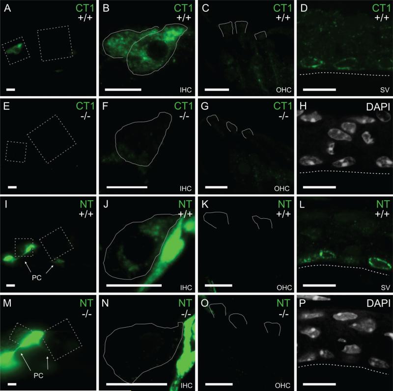
Figure 3.
TRPML3 redistribution to perinuclear vesicles of marginal strial cells and to inner hair cells of mature cochleae. A-C,E-G,I-K,M-O: Immunoreactivities on sections of the organ of Corti from juvenile (P29) mice with an antibody raised against a carboxyl-terminal domain of TRPML3 (CT1; A-C,E-G) and an antibody raised against an amino-terminal domain of TRPML3 (NT; I-K,M-O). Immunoreactivities were used with the same conditions as for neonatal inner ears (Fig. 2) or, to facilitate tissue adhesion to the slide, with milder conditions (shown here). Both antibodies label inner hair cells (IHCs; A,B,I,J), but not outer hair cells (OHCs; C,K) from Trpml3+/+ mice. These immunoreactivities likely represent TRPML3 protein, because they are not detected on sections from Trpml3−/− mice (E-G,M-O). As with the neonatal organ of Corti, the NT antibody labels pillar cells (PC) nonspecifically in both Trpml3+/+ (I,J) and Trpml3−/− (M,N) inner ears. D,H,L,P: Immunoreactivities on sections of adult (P147) stria vascularis with the same two antibodies (CT1 in D and NT in L) reveal that TRPML3 redistributed to vesicles surrounding the nuclei (labeled with DAPI in H and P) of marginal cells. Tissue from Trpml3−/− mice lacked these immunoreactivities. All pairs of Trpml3+/+ and Trpml3−/− images were acquired with same exposure, laser, and gain settings. The brightness and contrast settings were adjusted identically for each pair of images. Dashed boxes in A,E,I,and M indicate areas magnified to show IHCs and OHCs. White lines delineate full IHCs or apical regions of OHCs as determined by LAMP1 immunoreactivity (not shown). Dashed lines indicate apical side of marginal cells in stria vascularis. Scale bars = 10 μm. [Color figure can be viewed in the online issue, which is available at wileyonlinelibrary.com.]

