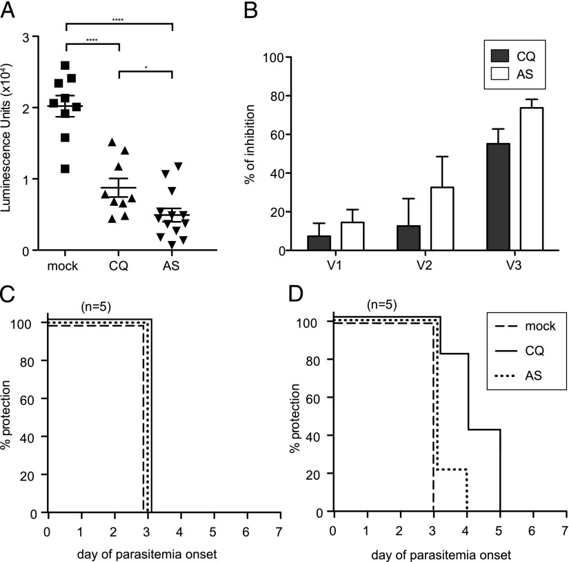
FIGURE 6.
Sera from mice treated with CQ- or AS-ITV inhibits spz invasion of hepatocytes in vitro. (A) Quantification of HepG2 cell invasion and development after incubation of P. yoelii GFP-luciferase spz with serum collected from mock-immunized, untreated (n = 9), ITV-CQ–treated (n = 9), or ITV-AS–treated (n = 13) mice 9 d after the third dose. The y-axis shows luminescence units. Significance was determined by two-tailed Student t tests. Results are from two independent experiments. Error bars represent SEM. *p < 0.05, ****p < 0.001. (B) Percent inhibition of HepG2 cell invasion and development upon incubation of P. yoelii GFP-luciferase spz with sera obtained after the first (V1), second (V2), or third (V3) course of CQ- or AS-ITV (AS), calculated as the percentage reduction in luciferase signal compared with that of the mock-immunized, untreated control. (C and D) Patency curves of naive mice challenged with wt spz (C) or iRBC (D) after adoptive transfer of immune sera from CQ-ITV–treated (solid line) or AS-ITV–treated (dotted line), or untreated, mock-immunized mice (dashed line). The y-axis shows the percentage of mice protected from infection at each time point, and the x-axis corresponds to the day of parasitemia onset after challenge.

