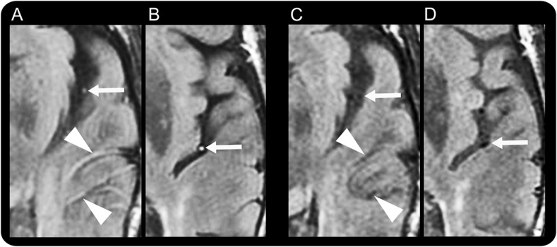
Figure. Vascular intensity changes on fluid-attenuated inversion recovery during and after resolution of symptoms in our TIA patient.

Fluid-attenuated inversion recovery vascular hyperintensities (FVH) during aphasia, including dot-like (arrows in A and B) and serpentine (arrowheads in A) hyperintensities, were seen in the middle cerebral artery branches. There were no abnormalities on diffusion-weighted or gradient echo images or magnetic resonance angiography. After aphasia resolution, the MRI showed FVH disappearance (C, D).
