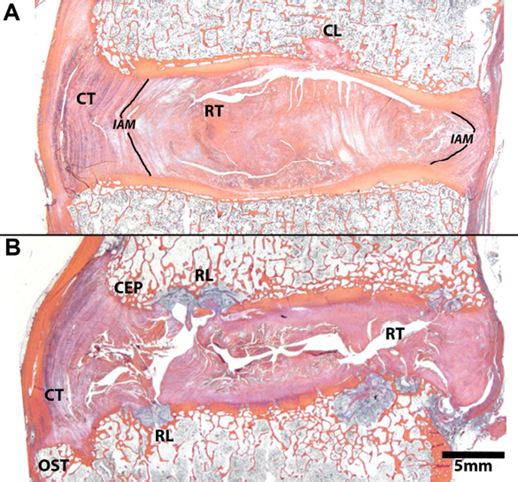
Fig. 3.
Mid-sagittal histological sections of human discs depicting histopathologies that coincide with disc degeneration. Both discs are Thompson grade 4 and from the same spine, the top image (A) is L4–L5 and the bottom image (B) is L5–S1. See Table 1 for definitions of labeled histopathologies. Note, the centrally-located lesion through the upper endplate in 3A labeled CL is a general endplate defect and is not by definition, a rim lesion. It differs from a rim lesion in its central location, adjacent to the nucleus pulposus. Histology sections in figure are stained with Mallory-Heidenhain.

