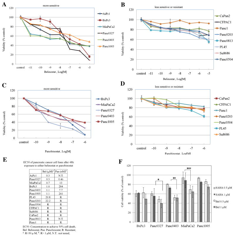
Figure 1.
Anti-proliferative activity of belinostat and panobinostat against 14 pancreatic cancer cell lines. Panels (A–D): MTT assays of pancreatic cancer cells cultured in 96-well plates and treated with drugs for 48 h. Cell numbers were measured after drug treatment relative to diluent controls. A series of dilutions (1 ×10⁻¹¹ M to 1 × 10⁻⁵ M) of belinostat (Panels A,B) or panobinostat (Panels C,D) were used. Panel (E): List of EC50s for belinostat and panobinostat against a panel of pancreatic cancer cell lines. Panel (F): Comparison between the anti-proliferative activity of belinostat and SAHA. Five pancreatic cancer cell lines were exposed to belinostat (0.5, 1 μM) or SAHA (0.5, 1 μM) for 48 h and MTT assays were performed. (*P = 0.07; **P = 0.013; ***P = 0.03). Results represent the mean ± SD of three independent experiments with quadruplicates at each time. Bel, belinostat; SAHA, veronistat.

