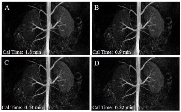
Figure 7.

A time resolved, contrast-enhanced, breath-held renal MRA exam was reconstructed using 3D through-time radial GRAPPA. This data was accelerated in-plane with a factor of 12.6 with respect to Nyquist, and a partial Fourier acquisition was used along the partition direction. The same coronal frame is shown here as MIPs for four different GRAPPA weight calibration schemes: 8 calibration partitions with 16, 8, 4, and 2 calibration repetitions (A, B, C, and D). Calibration acquisition times are noted in the lower left corner (Cal time). These images have a spatial resolution of 1.5mm × 1.5mm × 3mm and a temporal resolution of 3.5 s/frame.
