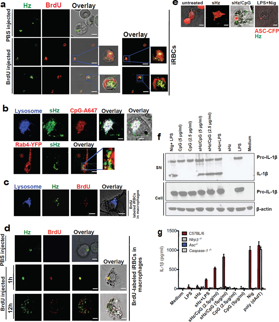
Figure 1. Hemozoin colocalizes with DNA in vivo is internalized into the phagolysosome where it has the capacity to prime macrophages and activate the NLRP3 inflammasome.
(a)Erythrocytes from PBS injected and BrdU injected P. berghei ANKA infected mice were stained with FITC-conjugated anti-BrdU antibody and subjected to confocal microscopy. Boxed areas in middle and bottom panels are enlarged (3×). Scale bar: 3µm (top panel), 7.8µm (middle panel) and 7.8µm (bottom panel). (b) Top: sHz (100µg/ml) was incubated with CpG–Alexa 647 (5µg/ml) for 2h. The complex was washed 3× with PBS before incubation with immortalized BMDMs and imaged by confocal microscopy after 30 min. Scale bar: 5µm. Bottom: Confocal microscopy of immortalized BMDMs stably transduced with Rab4-YFP, stimulated with sHz crystals (100µg/ml) for 2h. scale bar: 15µm. The boxed areas are enlarged on the right, demonstrating lack of co localization. (c) RBC from BrdU injected P. berghei ANKA infected mice were stained and prepared as in Fig. 1a. BMDMs were incubated with iRBCs (MOI: 5 to 1) × 1h and stained with lysotracker. Scale bar: 5µm. (d) RBC from PBS- or BrdU-injected P. berghei ANKA infected mice were incubated with BMDMs (MOI: 5 to 1), stained with FITC-conjugated anti-BrdU antibody and subjected to confocal microscopy after 1 or 12h. Scale bar: 20µm (top panel), 20µm (bottom panel). (e) BMDMs stably expressing ASC-CFP were treated with sHz/CpG (5µg/ml), sHz only (100µg/ml) or LPS (100ng/ml) × 2 h and then treated with nigericin (10µM), a K+ ionophore that activates NRLP3. The formation of ASC pyroptosomes was visualized by confocal microscopy. Fields are representative of at least 10 fields of view and 3 independent experiments. Scale bars from left to right: 15, 10, 5 and 5 µm; sHz (green) was visualized using reflection microscopy. (f) Immunoblot analysis of IL-1β cleavage to mature IL-1β (p17) in supernatants (SN) and cell extracts (Cell) from wt murine BMDMs stimulated as described in (g). (g) ELISA of released IL-1β by immortalized BMDMs from wt, Nlrp3−/−, Asc−/− and casp1−/− macrophages which were unprimed or primed or with LPS (100ng/ml) for 2 h, and then left unstimulated or stimulated for an additional 12 h with sHz, sHz/CpG (sHz: 100µg/ml and CpG: 2.5µg/ml and 5µg/ml), CpG (2.5µg/ml and 5µg/ml) or transfected with 1.5 µg/ml poly (dAdT). Nigericin (Nig) was used as a control. Poly (dAdT) was used as an AIM2 inflammasome activating control. Data are presented as mean ± SD of triplicates and are representative of 3 independent experiments. See also Fig. S1.

