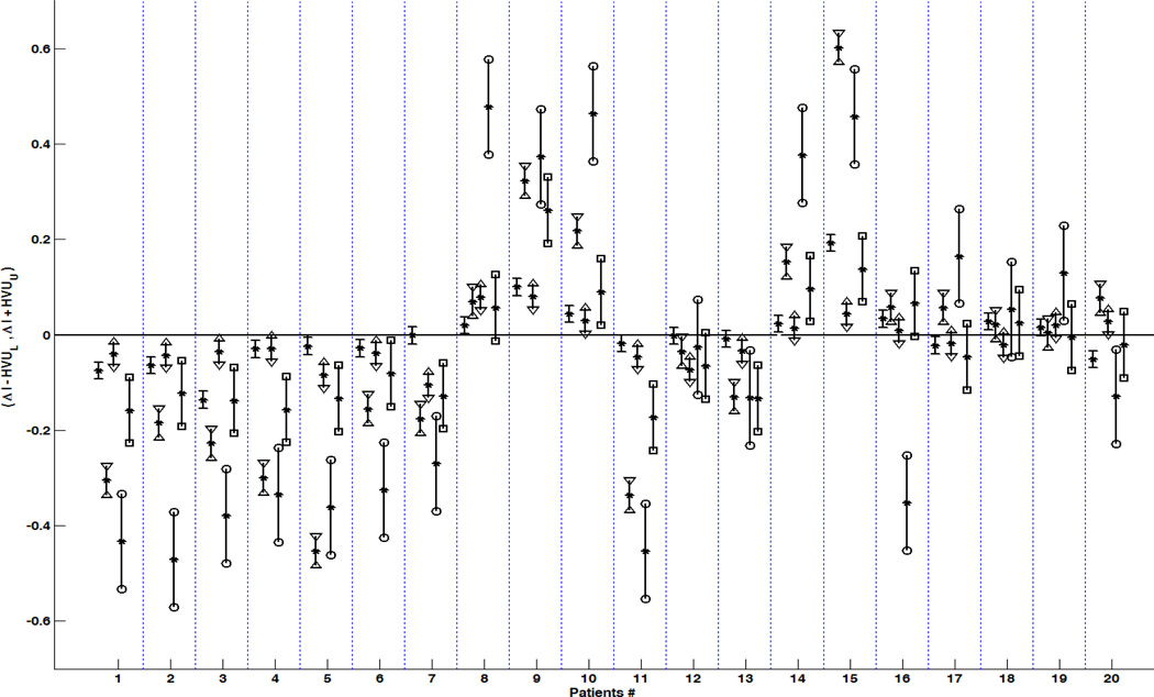
Figure 6. The 95% confidence interval (CI) of true hemispheric variations for individual TLE patients.
Suppose that μipsi and μcontra are true value of index I in ipsilateral and contralateral sides to epileptogenicity and ΔI = Iright − Ileft is the hemispheric variation (depicted by stars). The 95% CI of μcontra − μipsi was calculated as (ΔI − HVUU, ΔI + HVUU) for ΔI as the hemispheric variation of FA within the fornix crus (depicted between minuses), FA within the posteroinferior cingulum (depicted between triangles facing inward), the hemispheric variation of hippocampal MD (triangles facing outward), the hemispheric variation for hippocampal volumetrics (between squares), and the hemispheric variation for hippocampal FLAIR signal intensities (between circles). An individual patient was considered to have a true hemispheric variation if the 95% CI of the true hemispheric variation did not contain zero.
Note that the Patients# 1 to 10 belong to TLE patients with pathology proven MTS.

