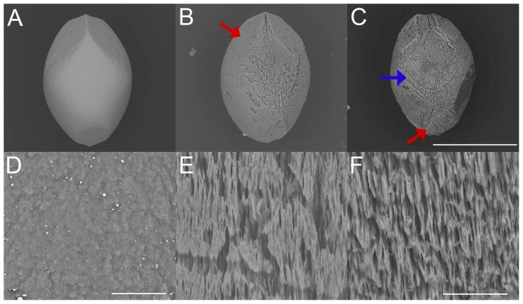
Figure 5. Dissolution of artificial otoconia and details of structural changes (belly-area), by treatment with demineralized water.
(A) Single artificial otoconium before treatment. Dissolution after 110 hours (B) and 200 hours (F) exposure. The red arrows in (B) and (C) show the position of one of the branches. The blue arrow in (C) points to the belly-area. (D) Surface of the belly-area before exposure and after 110 hours (E) and 200 hours (F) treatment. ESEM, low vacuum (LV), 15 kV. Scale bars (C) also for (A–B): 300 µm, (D): 10 µm, (F) also for (E): 20 µm.

