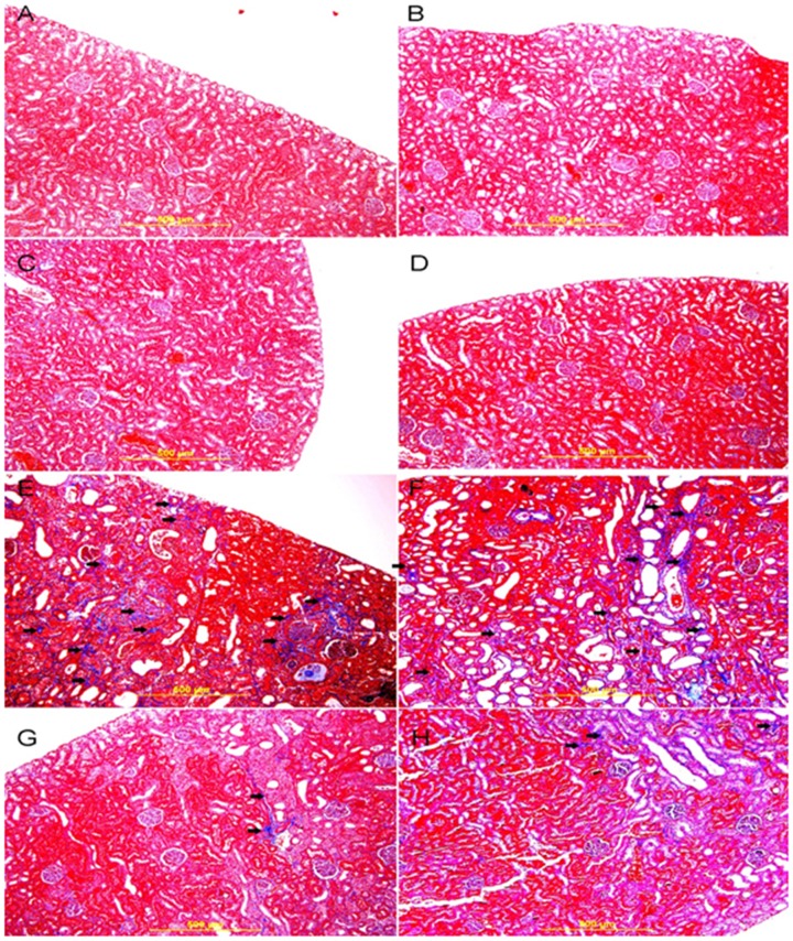
Figure 7. Representative photograph of sections of renal tissue of rats treated with saline (A), saline + swimming exercise, SE (B), gum acacia, GA (C), GA + SE (D), adenine (E), adenine + SE (F), adenine + GA (G) and adenine + GA + SE (H), and stained with Masson trichrome stain.
Sections A, B, C, and D showed normal kidney architecture and histology and no evidence of fibrosis. Sections E and F showed large areas of interstitial fibrosis (thick arrows). Sections G and F showed similar improvements in histological appearance with dramatic decrease in fibrosis (thick arrows).

