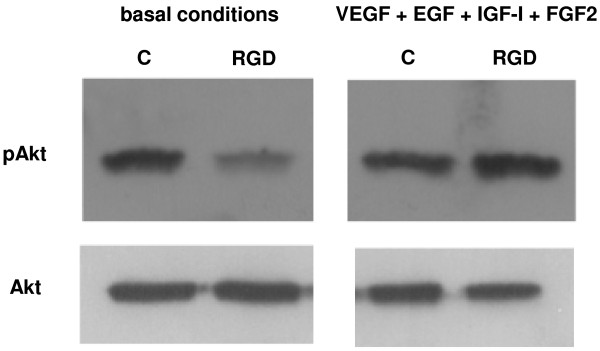
Figure 5.

Western blot analysis of Akt phosphorylation in HUVEC cultured for 5 h in basal conditions and with VEGF, EGF, IGF-I, and FGF2, alone (control, C) or in the presence of 1 μM cyclo[DKP-RGD] 1 (RGD). Data are from one representative of 3 separate experiments.
