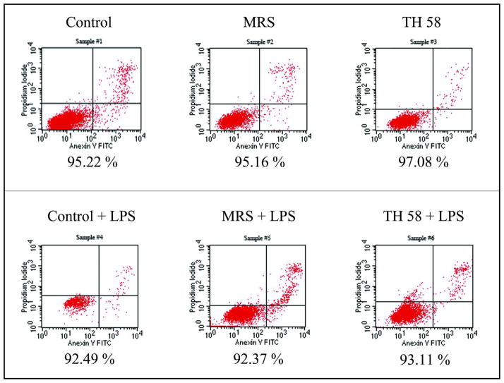
Fig. 3.
The viability of THP-1 cells determined by staining with Annexin V-FITC and Propidium Iodide (PI) followed by flow cytometric analysis. Viable cells are negative for both Annexin V-FITC and PI. Cells that are in early apoptosis are Annexin V-FITC positive and PI negative. Cells that are in late apoptosis or already dead are both Annexin V-FITC and PI positive. The percentages of cell viability under each plot are representative of three experiments. Control: RPMI cell culture medium; MRS: deMan-Rogosa-Sharpe, bacterial media control; TH58: Lactobacillus TH58 conditioned medium.

