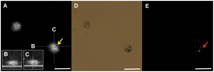
Figure 3. Representative image of cultured macrophage cell with cholesterol crystal.
A. Representative image of a macrophage with a cholesterol crystal. The macrophage on the right demonstrated highly scattering constituents inside its cytoplasm (yellow arrow). B, C. The Cross sectional images of the macrophage on the right. D, E. Polarization microscopy confirmed that the inclusions are cholesterol crystals (red arrow). Scale bars = 50 µm.

