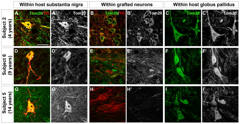
Figure 2. Mitochondrial phenotype in transplanted fetal dopamine neurons 4–14 years post-transplantation.
Double immunolabeling for translocase of outer mitochondrial membranes 20kDa (Tom20) (green) and tyrosine hydroxylase (TH) (red) in the host substantia nigra (left panels) and grafted dopamine neurons (middle panels) and in the host globus pallidus (right panels) in Subject 2 (panels A, A′, B, B′, C, C′, graft survival of 4 years), Subject 6 (panels D, D′, E, E′, F, F′, graft survival of 9 years) and Subject 5 (panels G, G′, H, H′, I, I′, graft survival of 14 years). Panels A′-I′ show single channel Tom20 labeling from corresponding panels A-I. In dopamine (TH-immunoreactive) neurons from the patients’ own substantia nigra, Tom20 labeling often appeared intensely labeled in the cell soma with accumulation in the perinuclear area (arrows) and little immunostaining in the dopaminergic axons and processes (panels A, A′, D, D′, G, G′). In grafted TH-immunoreactive neurons at 4 years post-transplantation Tom20 labeling was robust in the perikarya and neuronal processes (panels B, B′). At 9 and 14 years post-transplantation, Tom20 labeling was generally less intense in the grafted TH-immunoreactive neurons (panels E, E′, H, H′) compared to the Tom20 staining pattern observed in Subject 2 at 4 years post-transplantation, however, there was no abnormal accumulation of mitochondria in the cell soma as was observed in the host substantia nigra. No perinuclear accumulation or fragmentation of Tom20-labeled mitochondria was observed in the host globus pallidus (panels C, C′, F, F′, I, I′). Scale bar = 50μm

