Figure 2.
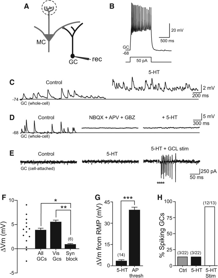
Indirect excitation of granule cells by serotonin. (A) Diagram of recording configuration. (B) Example granule cell (GC) response to a 50-pA depolarizing current step. (C) Example intracellular GC recording before and during 5-HT application. Horizontal line at −74 mV in both traces. (D) Blockade of fast synaptic responses with NBQX + APV + GBZ occludes depolarizing effect of 5-HT on GC. Horizontal line at −68 mV in all three traces. Mean membrane potential change in synaptic blocker cocktail was 0.4 ± 0.7 mV, n = 6, not significantly different from 0, P > 0.05. (E) Example cell-attached GC recording showing no spontaneous spikes before or after 5-HT application. Focal stimulation in the granule cell layer (GCL, asterisks) triggered a barrage of action currents in the same cell-attached recording (right trace). (F) Plot of change in mean intracellular membrane potential in 14 GCs treated with 200 µM 5-HT; ▵Vm from individual experiments indicated by symbols at left side. Serotonin triggered a similar membrane potential depolarization in subset of GCs with visualized and intact dendritic arbors within the external plexiform layer (Vis GCs, n = 4). Granule cell 5-HT response was greatly attenuated when fast synaptic responses were blocked by NBQX + APV + GBZ (Syn Block, n = 6). (**) P < 0.01, (*) P < 0.05. (G) Plot of mean GC depolarization triggered by 5-HT in control conditions (n = 14) relative to the membrane depolarization required to reach spike threshold from the same resting membrane potential (n = 14). (***) P < 0.001. (H) Plot of the proportion of GCs recorded in the cell-attached configuration which discharged spontaneously in control conditions (Ctrl, gray bar, 3/22), following bath application of 5-HT (3/22), and following focal GCL stimulation in 5-HT (12/13).

