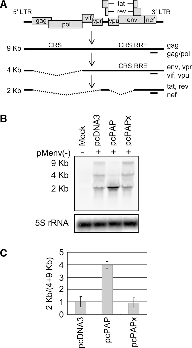
FIGURE 2.

PAP increases HIV-1 splicing and 2-kb mRNA accumulation. (A) Schematic representation of the HIV-1 genome organization, transcription, and splice products. Location of cis-repressive signals (CRS) and Rev responsive element (RRE) are indicated. The annealing site for the HIV-1-specific negative-strand riboprobe is indicated below each mRNA size class as a solid line. (B) 293T cells were cotransfected with pMenv(-) proviral clone (5 μg) and pcPAP, pcPAPx, or pcDNA3 (2.5 μg). Cells were harvested 40 h post transfection and Northern blotting was performed on total cellular RNA (15 μg). Levels of the three HIV-1 mRNA classes were visualized following hybridization with the HIV-1-specific probe. Blots were also hybridized with a 5S rRNA-specific probe as a loading control. (C) The abundance of HIV-1 mRNAs in each size class was measured by quantifying the band intensity using a PhosphorImager. The ratio of RRE-free mRNAs (2 kb) to RRE-containing mRNAs (9 and 4 kb) was plotted relative to 5S rRNA. Error bars represent means ± SE for three independent experiments.
