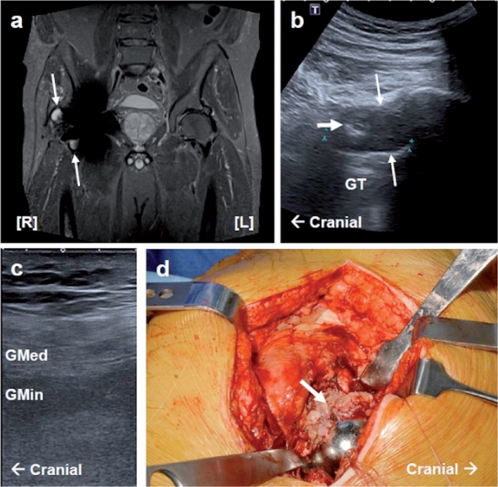
Figure 1.
Case 1. MARS MRI, ultrasound, and intraoperative images of a pseudotumor and gluteal muscle atrophy. a. A coronal STIR sequence MARS MRI section showing a right anterior (type-IIa) and lateral (type-IIb) pseudotumor (white arrows). In addition, right-sided fatty atrophy of the gluteus medius and minimus muscles (grade 3) can be seen. b. Lateral longitudinal USS showing a large cystic pseudotumor (type 2) with a thickened wall and upper solid focal region (thick white arrow). c. Lateral longitudinal USS of the right gluteus medius and minimus muscle showing fatty atrophy (reported as grade 2). d. Photograph taken during revision surgery showing a florid inflammatory reaction to the right hip neocapsule (thick white arrow).
GT: greater trochanter; Gmed: gluteus medius; Gmin: gluteus minimus. Pathology is indicated by white arrows.

