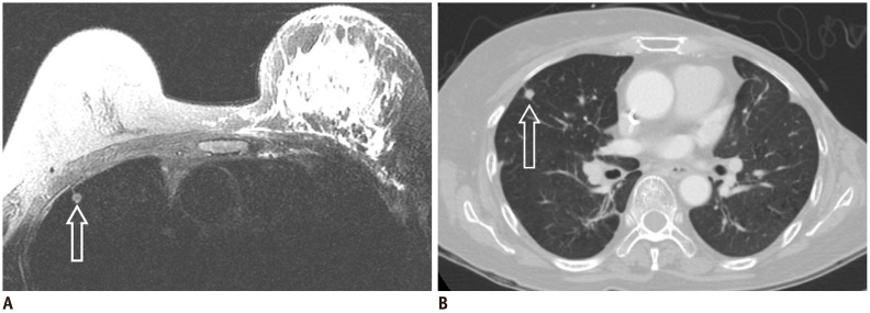
Fig. 1.
52-year-old female with left breast cancer with no changes in solitary pulmonary nodule on 24 months follow-up chest CT.
A. Inflammatory breast cancer was confirmed in left breast and solitary lung nodule was incidentally found in right upper lobe (hollow arrow) on T2-weighted image. B. Chest CT with lung setting shows solitary pulmonary nodule (hollow arrow) in right upper lung which shows no significant change 24 months after initial study (A).

