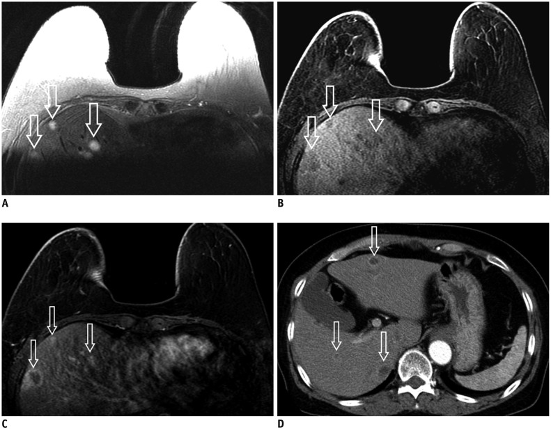
Fig. 8.
52-year-old female with right breast cancer and multiple hepatic metastases.
A-C. Liver contains several masses with indistinct margin (hollow arrows) with high signal intensity on T2-weighted image (A), low signal intensity on T1-weighted image (B) and nodular rim enhancement (hollow arrows) on contrast-enhanced T1-weighted image (C). D. Contrast-enhanced liver CT shows multiple peripheral rim-enhancing nodules (hollow arrows) in liver which are suggestive of hepatic metastases.

