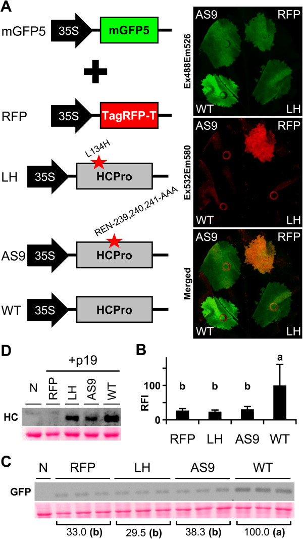
Figure 3.

Quantification of GFP accumulation in transient RNA silencing assay. (A) GFP was transiently expressed by co-infiltrating N. benthamiana leaf tissue with an Agrobacterium pBin-35S-mGFP5 culture plus cultures of Agrobacterium containing pSN.5 TagRFP-T (RFP), pSN.5 HC-L134H (LH, producing PPV HCPro L134H mutant), pSN.5 HC-AS9 (AS9, producing a PPV HCPro mutant in which amino acids REN-239, 240, 241 were replaced by alanines) or pSN.5 wtHC (WT, producing wild-type PPV HCPro). At 6 dpa, leaf fluorescence was acquired by laser scanning using Ex488/Em526 (green) and Ex532/Em580 (red); the image overlay is shown (Merged). (B) GFP fluorescence intensity of the agro-infiltrated leaf patches was quantified in a 96-well plate reader. RFI was plotted using WT mean value equal to 100. Bar graph shows mean ± SD (n = 14 biological replicates from two independent Agrobacterium cultures); the difference between the results marked with different letters is statistically significant, p < 0.01, one-way Anova and Tukey’s HSD test. (C) GFP protein accumulation in infiltrated leaves at 6 dpa was assessed by immunoblot analysis. Relative GFP signal intensities are indicated using average WT equal to 100; the difference between the values marked with different letters is statistically significant, p < 0.01, one-way Anova and Tukey’s HSD test. Each lane represents a pool of 3 or 4 leaf samples infiltrated with two independent Agrobacterium cultures. N, non-treated leaf sample. Ponceau red-stained blot as loading control. (D) HCPro expression by the binary vectors tested was assessed by HCPro immunoblot analysis of leaf co-infiltrated with an Agrobacterium pSN.5 p19 culture (6 dpa). Each lane represents a pool of infiltrated leaf samples. N, non-treated leaf sample. Ponceau red-stained blot as loading control.
