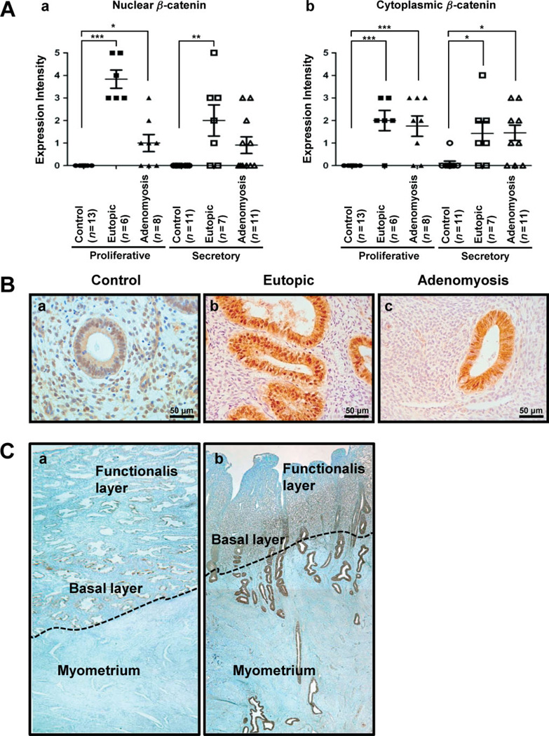
Figure 1.
Activation of β-catenin in endometrial tissue from women with adenomyosis. (A) Nuclear and cytoplasmic β-catenin was scored by measuring expression intensity of endometrial epithelial cells from control women (n = 24), eutopic endometrium (n = 13) and adenomyosis lesion (n = 19). (B) Photomicrographs represent immunostaining for β-catenin in human endometrium, with and without adenomyosis. β-Catenin was confined to the cell cytoplasm. Nuclear accumulation of β-catenin was prominent in eutopic endometrium (Bb, Bc). (C) The expression of β-catenin was observed in full-thickness endometrium, without (a) and with adenomyosis (b). ***p < 0.001; **p < 0.01; *p < 0.05; one-way analysis of variance (ANOVA) followed by Tukey’s post hoc multiple range test

