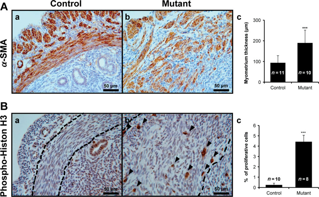
Figure 3.
Abnormal irregular structure and highly active proliferation in myometrium of uteri of mutant mice. (A) Immunohistochemical localization for smooth muscle actin (α-SMA), a smooth muscle cell marker, was examined in uterus of control (a) and mutant (b). The myometrial area of mutant mice were significantly increased (c) and revealed abnormal irregular structure. (B) Phospho-histone H3 was used for identification of proliferation in control (a) and mutant mice (b). Proliferative ability was increased in the myometrium of mutant mice compared with control mice (c); arrowheads, positive phospho-histone H3 cells; dashed lines, inner circular layer of myometrium. ***p < 0.001; one-way analysis of variance (ANOVA) followed by Tukey’s post hoc multiple range test

