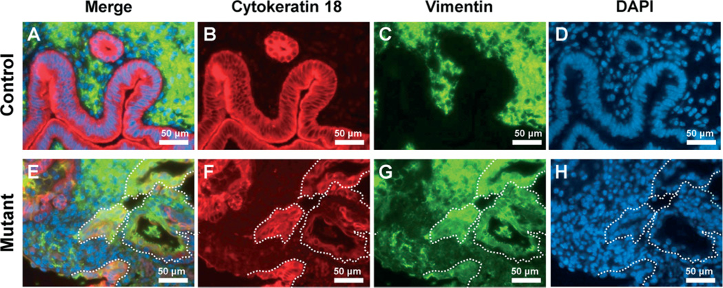
Figure 7.
The expression of vimentin in uteri of control and mutant mice. Immunofluorescence analysis of cytokeratin 18 (red; B, F) and vimentin (green; C, G) was performed in control (A–D) and mutant (E–H) mice. The expression of vimentin was observed in epithelial cells of the uterus in mutant but not control mice; white dotted lines, vimentin-positive epithelial cells

