Figure 8.
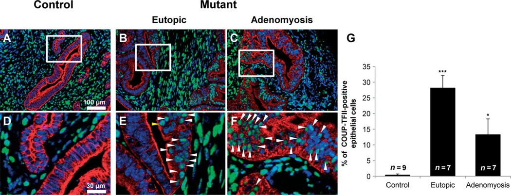
The expression of COUP-TFII in uteri of control and mutant mice. Immunofluorescence analysis of COUP-TFII (green) and cytokeratin 18 (red) was performed in uteri of control (A, D) and mutant (B, C, E, F) mice. COUP-TFII was observed in epithelial cells of mutant mice (B, C, E, F), while its expression was limited to endometrial stromal cells and the myometrium of control mice (A, D). (D–F) High-magnification pictures of the boxed areas in (A–C); arrowheads, COUP-TFII-positive cells. (G) COUP-TFII-positive cells were significantly increased in mutant mice. *p < 0.05; ***p < 0.001, one-way analysis of variance (ANOVA) followed by Tukey’s post hoc multiple range test

