
Fig 1. Loss of NPM1 results in more constitutive and IR-induced γH2AX foci.
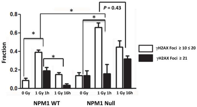
NPM1-wt and NPM1-null MEFs were administered 0 or 1 Gy. Irradiated cells were incubated at 37°C for 1 or 6 hrs. Histogram showing the fraction of cells with 11–20 (white bars) or >20 (black bars) γH2AX foci N= 200–250 nuclei per point. *P < 10−3 2 tailed Fisher’s Exact Test.