
Fig 2. Rad51 foci formation.
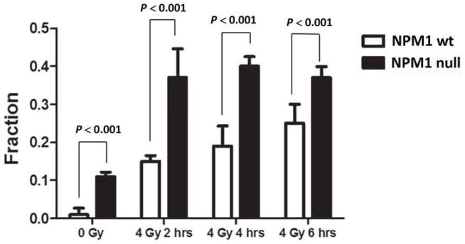
NPM1-wt and NPM1-null MEFs were administered 0 or 4 Gy. Irradiated cells were incubated at 37°C for 1–6 hrs. Histogram showing the fraction of nuclei with greater than 5 Rad51 foci as a function of time after 4Gy irradiation. N=100 nuclei per point. The results represent the average (error bars: +/− SD) from three independent experiments.