Figure 2.

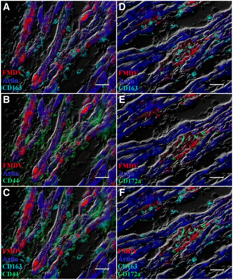
Myocarditis in two pigs infected with FMDV A24 Cruzeiro (case 1; A-C) or FMDV O1 Campos (case 2; D-F). In both cases, FMDV VP1 capsid protein co-localizes with actin in cardiomyocytes. Myocardium from case 1 contains marked infiltrates of overlapping populations of CD44+ and CD163+ cells (B-C). Myocardium from case 2 has low numbers of CD172a + and CD163+ cells (E-F). A-C: FMDV A capsid (red), actin (blue), CD163 (turquoise), CD44 (green). D-F: FMDV O capsid (red), actin (blue), CD163 (turquoise), CD 172a (green). Multichannel immunofluorescence. Scale bars = 25 μm.
