Figure 2.

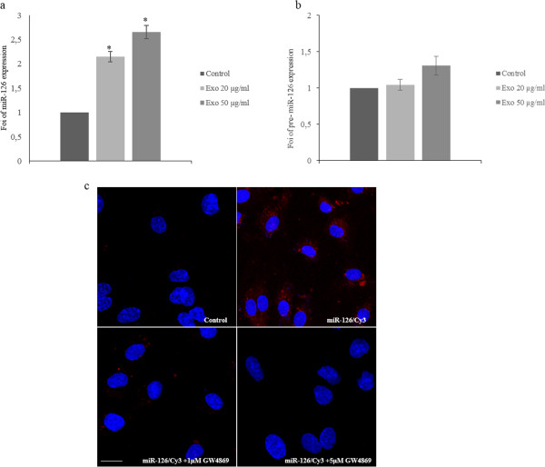
Exosomes shuttle miR-126 in HUVECs. a: MiR-126 expression in HUVECs treated with different amounts of LAMA84 exosomes. miR-126 expression levels in HUVECs treated with 20 and 50 μg/ml of LAMA84 exosomes for 24 hours were determined by quantitative Real time PCR analysis. Values are the mean ± SD of 3 independent experiments *p ≤ 0.05. b: Pre-miR-126 expression in HUVECs treated with different amounts of LAMA84 exosomes. Pre-miR-126 expression levels in HUVECs treated with 20 and 50 μg/ml of LAMA84 exosomes for 24 hours were determined by quantitative Real time PCR analysis. c: Localization of exosomal miR-126 into HUVECs. HUVECs were co-cultured with LAMA84/Cy3-miR-126 cells using Transwells. In Red is shown Cy3-miR-126 in the cytoplasm of HUVECs (miR-126/Cy3), nuclear counterstaining was done with Hoescht (blue). As a negative control, HUVECs were co-cultured with untrasfected LAMA84 (Control). HUVECs were also cocoltured with LAMA84/Cy3-miR-126 cells treated with 1 μM (miR-126/Cy3 + 1 μM GW4869) and 5 μM (miR-126/Cy3 + 5 μM GW4869) of GW4869.
