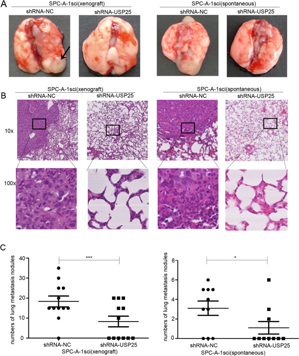
Figure 6.

The promoter roles of USP25 on lung metastasis in vivo. (A, B) Representative gross photo of a mouse lung and the images of histological inspection of a mouse lung for the presence of microscopic lesions eight weeks after tail vein injection (xenograft) or subcutaneous injection (subcutaneous) with SPC-A-1sci cells stably expressing the sh-USP25 or sh-NC. (C) Quantification of lung microscopic nodules in the lungs of each group. Statistical analysis was performed using Student’s t-test. Error bars represent S.E.M. *P < 0.05; **P < 0.01; ***P < 0.001.
