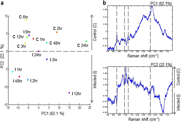
Figure 5.

PCA discrimination of medium from healthy and Neospora caninum-infected cell culture media with respect to time post infection. Panel a depicts the PCA for time-resolved Raman spectral data, score plot of axes 1 and 2. Principal components 1 and 2 explained 62.1and 22.1% of the variance in the data, respectively. Samples at different time points are indicated by different colours. Panel b depicts the loadings plot for the analysis shown in panel A showing the discrimination between the spectra of control (C) and (I) infected culture media. The dotted lines in loading plots mark the location of the Raman bands depicted in Figure 4.
