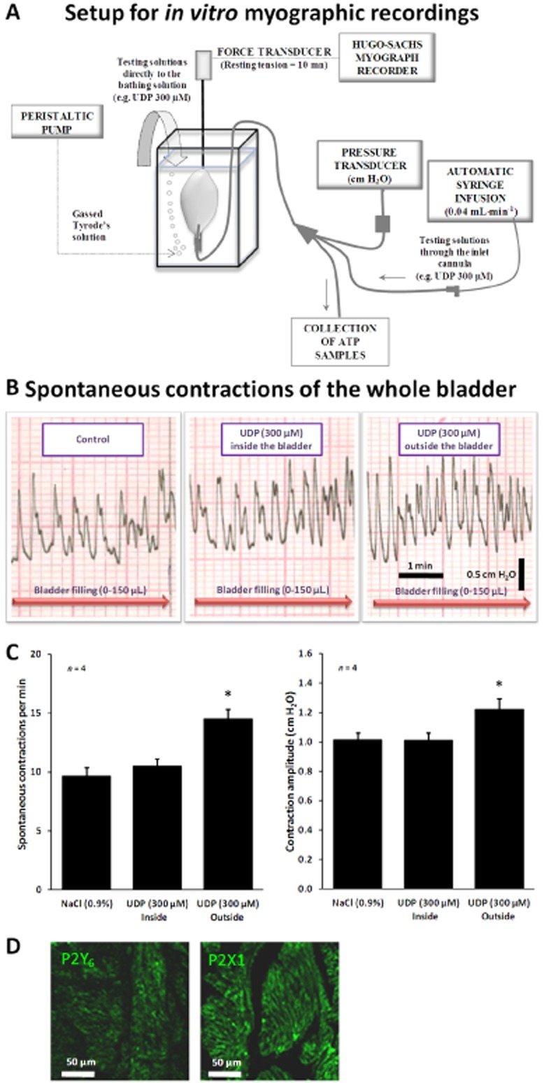
Figure 6.

(A) Setup for myographic recordings of the whole urinary bladder of the rat in vitro. (B) Spontaneous contractile activity of the rat urinary bladder in response to bladder filling with Tyrode's solution, up to 150 μL, infused at a constant flow rate (40 μL·min−1) to mimic in vivo cystometry experiments. UDP (300 μM) was superfused either into the bladder lumen (by changing the syringe connected to the automated perfusion system) or directly to the bathing solution outside the bladder. (C) Quantification of the frequency and magnitude of spontaneous contractions of the whole bladder in vitro in the absence and in the presence of UDP (300 μM) applied inside and outside the bladder. The vertical bars represent SEM of four isolated bladders. *P < 0.05; significantly different from control (saline superfusion); one-way anova followed by Dunnett's modified t-test. (D) Confocal micrographs of transverse sections of rat urinary bladder detrusor muscle immunostained for P2Y6 (APR-011) and P2X1 (APR-001) receptors. Scale bars = 50 μm.
