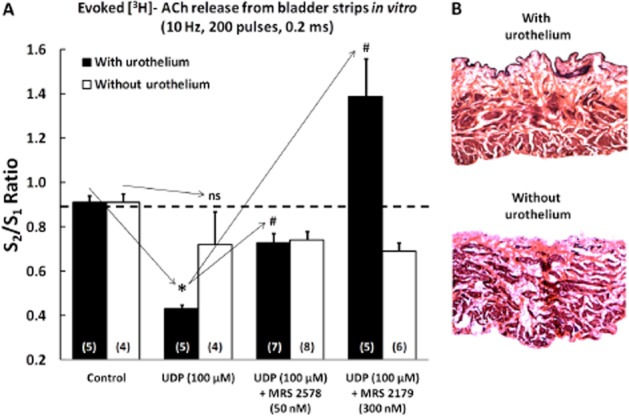
Figure 8.

(A) Effect of UDP (100 μM) on electrically-evoked [3H]-ACh release from intact urinary bladder strips and in preparations without the urothelium UDP (100 μM) was applied 8 min before S2. MRS2578 (50 nM) and MRS2179 (300 nM) were added to the incubation media at the beginning of the release period (time zero) and were present throughout the assay, including S1 and S2. The ordinates represent evoked tritium outflow expressed by S2/S1 ratios, i.e. the ratio between the evoked [3H]-ACh release during the second period of stimulation (in the presence of UDP) and the evoked [3H]-ACh release during the first stimulation period (without UDP). The vertical bars represent SEM. *P < 0.05; significantly different from control; #P < 0.05: significantly different from UDP alone; unpaired Student's t-test with Welch's correction. (B) Representative microscopic images of rat urinary bladder strips stained with haematoxylin-eosin to confirm the presence or the absence of the urothelium. Magnification, 40×.
